Scientists have long wondered why sleep is restorative and why lack of sleep impairs brain function.
Now, new animal research suggests how the sleep state may help clear the body of potentially toxic central nervous system (CNS) metabolites.
Proteins linked to neurodegenerative diseases, including β-amyloid (Aβ), are present in the interstitial space surrounding cells in the brain. In a series of experiments, researchers tested the hypothesis that Aβ clearance is increased during sleep and that the sleep-wake cycle regulates the glial cell–dependent glymphatic system, which is responsible for clearing waste from the brain and spinal cord.
"Basically, we found a new function of sleep," said study lead author Lulu Xie, PhD, Division of Glial Disease and Therapeutics, Center for Translational Neuromedicine, Department of Neurosurgery, University of Rochester Medical Center, New York.
"When mice are awake, the brain cells continuously produce toxic waste. This waste can build up in the spaces between the brain cells and damage them. However, during sleep, the spaces between brain cells increase, which may help the brain flush out the toxic waste. Therefore, a good sleep can clear the brain."
"Sleep changes the cellular structure of the brain. It appears to be a completely different state," Maiken Nedergaard, MD, DMSc, codirector of the Center for Translational Neuromedicine at the University of Rochester Medical Center, who is a leader of the study, said in a statement from the National Institute of Neurological Disorders and Stroke, which supported the study.
The new research was published October 18 in Science.
Sleeping vs Awake Brain
The researchers infused fluorescent dye into the cerebrospinal fluid (CSF) of mice and observed it flow through the brain. At the same time, they monitored electrical brain activity and wakefulness with electrocorticography (EcoG) and electromyography (EMG).
Dye flows through the brain of a sleeping mouse.Courtesy of Nedergaard Lab, University of Rochester Medical Center.
|
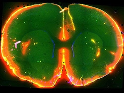
"In the sleeping brain, we found the CSF flushed into the brain very quickly and broadly," said Dr. Xie. "After half an hour, we woke the mice up by gently touching their tails, and injected another color of dye. But what we saw is that CSF barely flowed when the same mice were awake."
These results suggest that the awake brain may have more resistance to CSF influx, which leads to the assumption that the path of CSF flow into the brain is smaller in the awake brain, said Dr. Xie.
Next, the scientists inserted electrodes into the brain to directly measure the space between brain cells, and found that it increased by around 60% when the mice were asleep.
"Theoretically, big spaces lead to easier fluid influx," said Dr. Xie. "So we presumed that the clearance of the toxic protein between cells will become more efficient."
To test this assumption, they infused radio-labeled Aβ into the brain and measured how long it stayed in both the sleeping brain and the awake brain.
"We found Aβ disappeared 2-fold faster in the sleeping mice brains as compared with awake mice," noted Dr. Xie. "Based on this experiment, we can see that the sleeping brain is more capable of clearing out the toxic protein."
Technically, it might be relatively easy to study these processes in humans, possibly using magnetic resonance imaging. However, Dr. Xie said she does not know when human trials, which involve "a lot more concerns" than animal experiments, might come about.
"These results may have broad implications for multiple neurological disorders," said Jim Koenig, PhD, a program director at the National Institute of Neurological Disorders and Stroke (NINDS), which funded the study, in a statement. "This means the cells regulating the glymphatic system may be new targets for treating a range of disorders."
The study was funded by grants from the NINDS.
Science. 2013;342:373-377. Abstract
No comments:
Post a Comment